
х
すべてのiLiveコンテンツは、可能な限り事実上の正確さを保証するために医学的にレビューまたは事実確認されています。
厳格な調達ガイドラインがあり、評判の良いメディアサイト、学術研究機関、そして可能であれば医学的に査読された研究のみにリンクしています。 かっこ内の数字([1]、[2]など)は、これらの研究へのクリック可能なリンクです。
当社のコンテンツのいずれかが不正確、期限切れ、またはその他の疑問があると思われる場合は、それを選択してCtrl + Enterキーを押してください。
カリシウイルス
記事の医療専門家

アレクセイ・クリヴェンコ、医療評論家
最後に見直したもの: 08.07.2025
最後に見直したもの: 08.07.2025
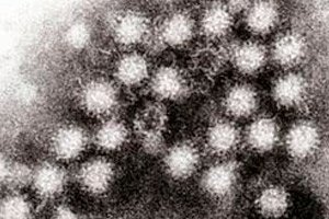 ">
">カリシウイルスは1932年に初めて動物から分離され、1976年に急性胃腸炎を患う子供の糞便から発見されました。現在では、カリシウイルス科という別の科に分類されています。
ウイルス粒子は球形で、直径37nm、スーパーカプシドは存在しません。ゲノムは分子量約2.6~2.8MDのプラス鎖一本鎖RNAで構成されています。ネガコントラスト顕微鏡観察では、ウイルス粒子の表面に32個の深い(約10nm)カップ状の窪みが認められ、これがカリシウイルス(ギリシャ語の「calyx(カップ)」に由来)と命名された理由です。カリシウイルスは細胞培養では増殖しないため、検出が困難です。診断には主に免疫電子顕微鏡法が用いられます。
 [ 1 ], [ 2 ], [ 3 ], [ 4 ], [ 5 ], [ 6 ], [ 7 ], [ 8 ], [ 9 ]